
Axon Structure and Dynamics
[Sub topic of Disease Process of ALS]
 |
|
When materials cannot move along the neuron to the muscle both cells suffer |
Active transport along the extensive axons of motor neurons conveys newly made materials to even the farthest reaching nerve endings, and needed nutrients back to the cell body. Motor neurons may be particularly vulnerable to any genetic defect or cellular insult that impedes axon transport. Studies of the proteins within nerve fibers have highlighted their importance in maintaining motor neurons, and point to the possible role of these axon proteins in ALS.
What are the Dynamics of Neurons Implicated in ALS?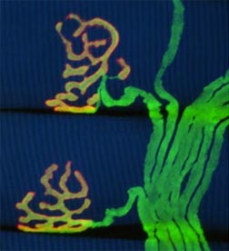 |
| Misgeld and Lichtman; neuromuscular junction in a transgenic mouse expressing YFP in motor axons, red: fluorescently labelled acetylcholine receptors in the muscle membrane. |
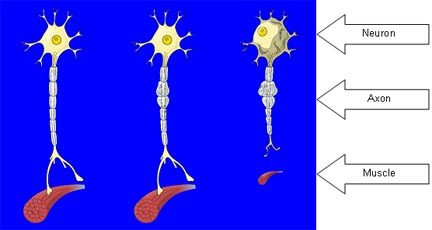
In ALS, the progressive weakness that develops is due to selective death of motor neurons, the nerve cells that carry messages telling the muscles to contract. Motor neurons are unique cells, the longest in the body. All neurons extend a connecting fiber, the axon, to the next neuron or end organ. Some motor neurons in the spinal cord must extend their axon up to a meter, to reach the toes, for example, yet the cell body maintaining this extraordinary fiber is only of ordinary size. Metabolic demands on the motor neurons must be correspondingly extraordinary.
All axons contain molecules within that serve as a scaffold. Axons also have a variety of proteins that serve as molecular ferries to shuttle cellular supplies. Active transport along the extensive axons of motor neurons conveys newly synthesized material from the center of the cell to the nerve endings, and also moves growth promoting factors from the endings back to the cell body. Axonal transport is mandatory for normal neuronal function. And motor neurons may be particularly vulnerable to genetic defects or cellular insults that impede axonal transport.
Crucial Nerve Proteins
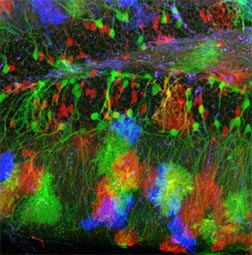 |
| Livet, Sanes, and Lichtman; a "Brainbow" transgenic mouse expressing three fluorescent proteins in different neurons and astrocytes in the hippocampus. |
Neurofilaments are proteins within neurons, key to defining and maintaining the structure of the axon. Scientists have shown that disruptions in the neurofilaments can induce these proteins to accumulate, and this can damage the motor neurons to produce symptoms bearing many similarities to ALS.
Tubulins are the building blocks of microtubules, another component of the axon’s scaffold. Researchers have found several mutations in proteins associated with microtubules that produce motor neuron disease. These findings highlight the important role of microtubules in maintaining motor neurons, and point to a possible role in ALS.
Dynein is still another component of the axon. It acts like a molecular motor for returning needed materials from the nerve endings back to the cell body. Transport from the neuromuscular junction back to the cell body involves both the motor protein dynein and its activator dynactin. A mutation in dynein, and in dynactin, each produce a motor disorder with certain similarities to ALS, pointing to this axon protein as a potential target for ALS therapeutics.
Related recent news releasesJoint Project Brings Expertise to Bear on ALS Axon Dynamics
Protein’s Role Emerging in ALS and Cognitive Change
Axon dynamics workshop
ALSA-Funded Research Presented at Society for Neuroscience Annual Meeting
Related ALSA Funded Research Projects
Click here to search ALSA Funded Research Projects